Our Services
What you can expect
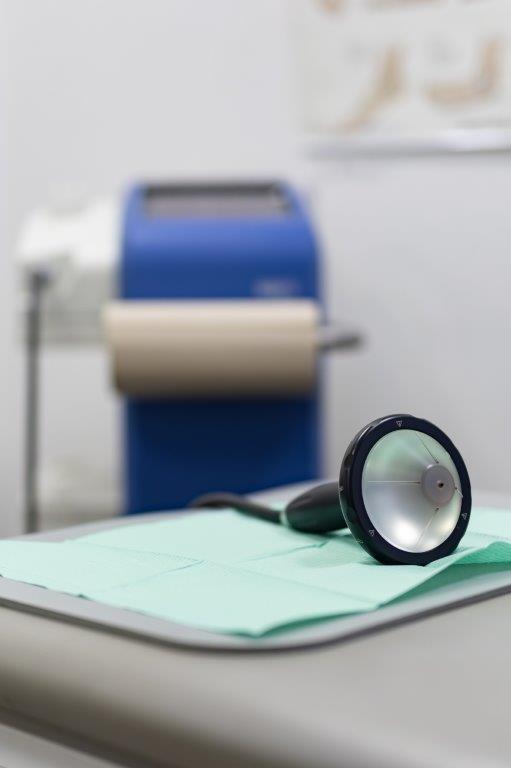
We offer the full spectrum of modern diagnostics for foot and ankle ailments, conservative treatments, and the very latest surgical techniques.
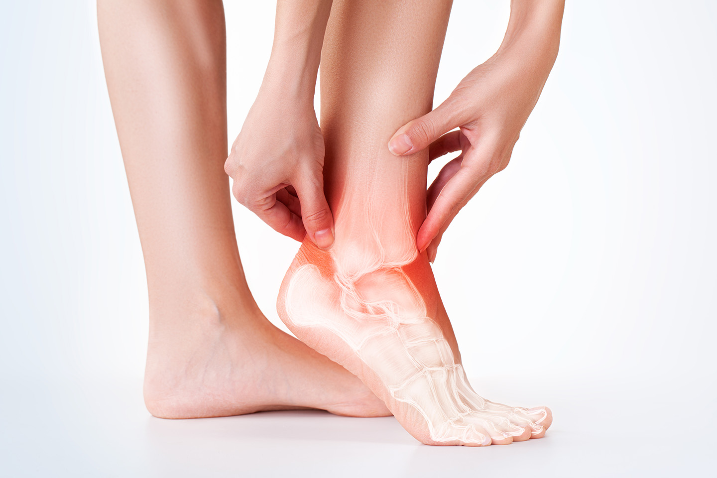
Diagnostics
Using state-of-the-art methods to identify the problem
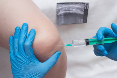
Ultrasound (sonography)
 Instrumental gait analysis
Dynamic pedography (dynamic plantar pressure distribution measurement)
Four-dimensional spine measurement
Electromyography (EMG, muscle function assessment), gastrocnemius soleus complex
Diagnostic radiology
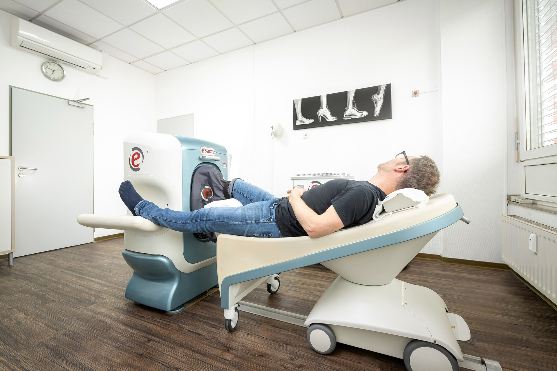
MRI (magnetic resonance imaging), open MRI
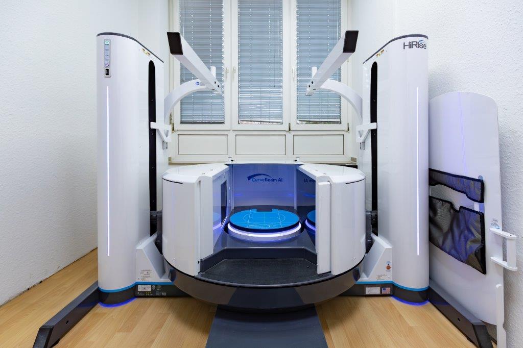
Digitale Volumen Tomographie (DVT) – 3D-Weightbearing-CT
Instrumental gait analysis
Dynamic pedography (dynamic plantar pressure distribution measurement)
Four-dimensional spine measurement
Electromyography (EMG, muscle function assessment), gastrocnemius soleus complex
Diagnostic radiology
MRI (magnetic resonance imaging), open MRI
Digitale Volumen Tomographie (DVT) – 3D-Weightbearing-CT
Ultrasound (sonography)
Ultrasound scans (sonography) allow us to view the feet and their surrounding structures (muscles, nerves, tendons). This lets us see whether fluid has collected in the ankles, tendons and ligaments have changed, or articular capsules (joint capsules) have become swollen. This makes it possible to diagnose inflammation more quickly, so that we can immediately begin treatment – such as for inflamed tendons or arthritis.Instrumental gait analysis
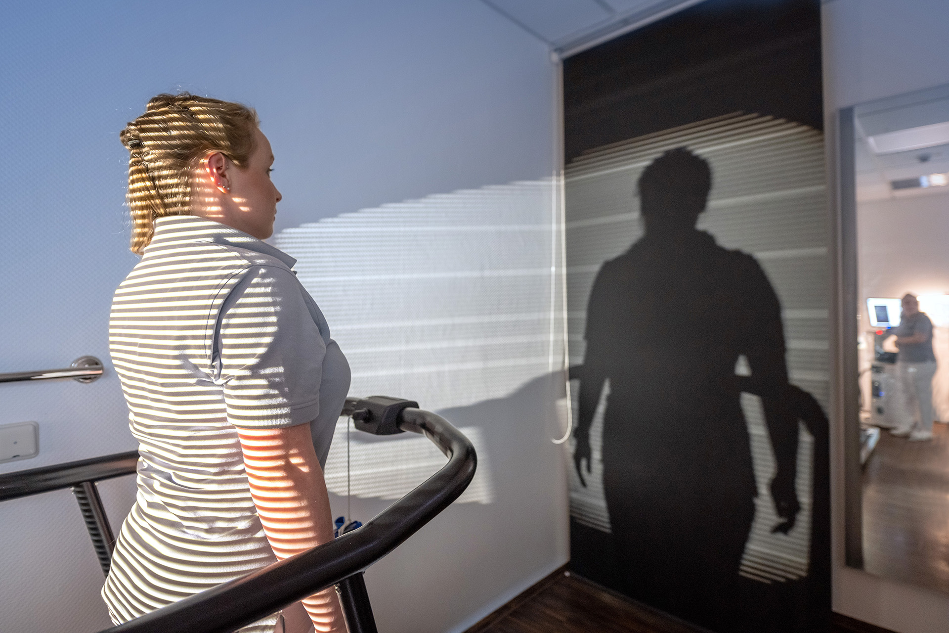
Instrumental gait analysis supplies us with precise, quantifiable data that allow us to measure the strain placed on feet and joints. We can recognise misalignments and improper gait, and initiate appropriate corrective measures and treatments.Dynamic pedography (dynamic plantar pressure distribution measurement)
We conduct a detailed analysis of the foot and precisely measure the pressure distribution and load on the foot using pedobarography (plantar pressure distribution measurement). These measurements supplement the instrumental gait analysis.Four-dimensional spine measurement
Four-dimensional spine measurement allows us to precisely record the position and shape of the spinal column and body. Deformities, malpositions, curvatures and distortions can be directly identified in this way. The examination takes place while the patient is standing and while running. Not only does this not involve any pain or radiation, but the detailed diagnostics enable us to begin initial treatment measures straight away.Electromyography (EMG, muscle function assessment), gastrocnemius soleus complex
We use EMG (electromyography) to examine muscle activity in order to assess muscle function and identify any disorders. With the EMG we focus on the gastrocnemius muscle, which is also known as the lower leg muscle or calf muscle. This muscle controls the bending and twisting of the foot, and allows us to flex our feet. As a result, this muscle plays an extremely important role when running, jumping and walking, as well as when riding a bicycle. We measure the muscle when fully and gently tensed, as well as when it is at rest, by sticking electrodes to the muscle that measure the tension. If the muscles or nerves are damaged, this will show up in the muscle activity.Diagnostic radiology
This imaging technology can reveal the presence of injuries or ailments, such as a bone fracture or damaged tissue. We also use X-rays to determine whether there might be sprains or dislocations, changes to the joints, osteoporosis, (bone loss), cartilage loss, or a tumour. Depending on the area being examined, the patient will sit or stand for this process, which takes just a few minutes.MRI (magnetic resonance imaging), open MRI
When we suspect inflammation of the muscles, tendons or joints, or when the other examination methods used have not been able to reveal the precise cause of pain being suffered, we recommend magnetic resonance imaging (MRI). MRI images are extremely precise, which makes it possible to provide reliable diagnoses. This is particularly useful for the proper recognition and depiction of stress reactions and foot fractures and allows us to commence suitable treatments quickly. The advantage of our open MRI: there is no need to feel claustrophobic. Our MRI machine is comfortable, convenient, quiet and safe, and it delivers high-resolution images of the precise areas being examined. Our practice holds the highest MRI certification awarded by the ADO (Academy of German Orthopaedists), the DGOU (German Society for Orthopaedics and Trauma Surgery), and the BVOU (German Professional Association for Orthopaedics and Trauma Surgery). This certifies us to perform and expertly evaluate musculoskeletal MRI examinations, ensuring precise, safe, and high-quality diagnostics.Digitale Volumen Tomographie (DVT) – 3D-Weightbearing-CT
State-of-the-art diagnostics under load
As one of the leading practices for foot and ankle surgery in Cologne, we now offer one of the most advanced imaging technologies in the world: the weightbearing CT (DVT under load). Only very few centers in Germany have a device capable of performing a complete examination of the entire foot, in some cases even both feet, knees, or hips at the same time. This allows us to precisely assess the leg axis up to the hips and use the opposite side as a reference for future corrections.
Unlike conventional CT or X-ray scans, the images are taken under real load – that is, while standing. This enables us to accurately evaluate the true position and function of your foot and ankle during everyday activity.
The result: three-dimensional, high-resolution images that reveal even the smallest changes in bones, joints, and soft tissues. This technology revolutionizes diagnostics and therapy, both conservative and surgical, and allows for more individualized and precise treatment decisions.
conservative treatments
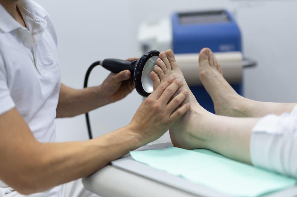
Focused ESWT (extracorporeal shock wave therapy)
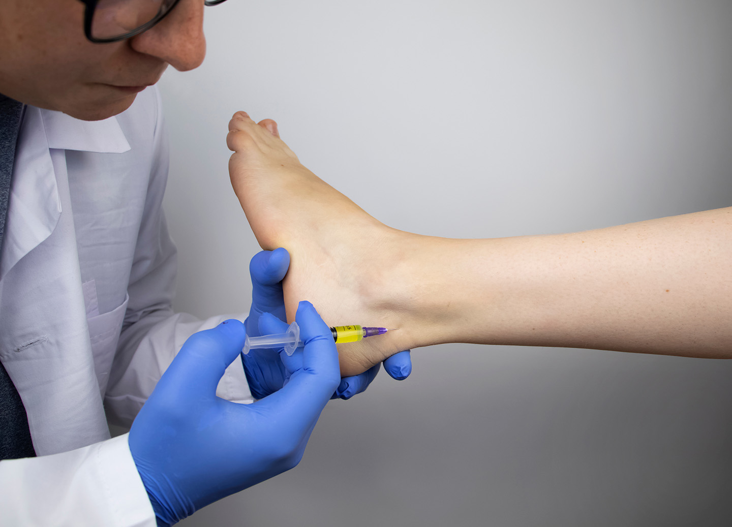
 Joint infiltrations (joint injections)
 Tendon injections
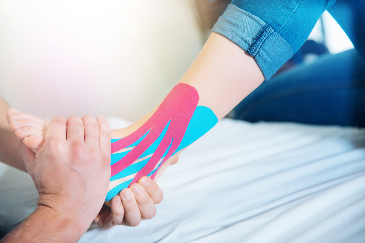
 Kinesio taping
 STW DIGEST
Focused ESWT (extracorporeal shock wave therapy)
We employ this procedure to treat chronic pain. With focused ESWT we can target the acoustic waves at a particular point, which makes it possible, for example, to optimise the stimulation of tissues located far below the skin. At the same time, these acoustic waves stimulate the nerve fibres that alleviate pain, with the result that the body releases anti-inflammatory substances. Tense muscles are relaxed, and blood circulation in the surrounding tissue is improved. We use ESWT for:- Heel spurs
- Delayed bone fracture healing
- Other bone fracture healing disorders
- Achilles tendonitis
- Painful inflammation of the tendon insertions
- Inflammation of the tendons or joints
Joint infiltrations (joint injections)
We provide you with this treatment if you are suffering from acute or chronic pain. This involves local injections that allow the medicine to work precisely in the affected area. Local cortisone therapy (anti-inflammatory agent) Cortisone is one of the body’s own hormones that controls numerous metabolic and immune system processes. Cortisone can help to combat severe inflammation, so it is also used for the treatment of arthritis. Hyaluronan (viscosupplementation) Hyaluronan is a substance produced naturally by the body that constitutes a significant part of joint fluid (synovial fluid). It works as a ‘joint lubricant’ and shock absorber. If the joint fluid does not contain enough hyaluronan, this can result in pain, improper joint and cartilage function, or damage to (and stiffness of) the joint. We inject hyaluronan into the ankle joint to alleviate the pain and stop symptoms from growing more severe. Autologous conditioned plasma (ACP) regenerative therapy This biological process is the very latest therapy for joint arthritis and inflammation of the tendons. The body’s own (i.e. autologous) plasma is extremely effective and easily tolerated by the body. It spurs the body to initiate its own healing process. We take your blood and process it to separate the plasma. We then inject the plasma into the affected location so that it can directly trigger the body’s own healing and regeneration processes. Even in cases of chronic tendonitis or inflammation of the tendon insertions, ACP therapy is effective – including for therapy-resistant cases of plantar fasciitis and Achilles tendonitis.
Tendon injections
We inject an anti-inflammatory agent into the tendon insertion in order to quickly put an end to acute pain, such as with tenosynovitis. An injection may be a one-off treatment, or it may need to be repeated more than once. An infiltration can break the pain cycle, and depending on the composition of the medication used, it may also be possible to treat the underlying ailment. Here, too, we can employ ACP regenerative therapy, as with joint infiltrations.
Kinesio taping
Kinesio tape can be used preventively or to treat symptoms. This tape can help the patient retain movement in the ankle joint. At the same time, its stretchability allows the tape to provide support for the foot. We use Kinesio tape for torn ankle ligaments, bunions, flatfoot/splayfoot, plantar fasciitis, Haglund’s heel, and when the upper ankle joint is unstable.
STW DIGEST
Our Center holds the full specialist certification for shockwave therapy, issued by the German-speaking International Society for Shockwave Therapy (DIGEST). We are the only center in Cologne that possesses this certification. DIGEST is the scientific association that defines the highest quality standards in this field. For you, this means precise indication and the best possible treatment results – ensuring a fast and lasting recovery. For more information, please visit the DIGEST website.
Our Specialities
Minimally invasive surgery (MIS)
Is an operation required to alleviate your symptoms? We devote our full expertise and the latest technology to your treatment – so that you will be back on your feet in no time and living life to the full.

Bunion treatment

Bunionette treatment

Hammer toe treatment

Achilles tendon disorders (Haglund’s heel)

Morton’s neuroma
Ankle Joint Replacement (Ankle Endoprosthesis)
Surgical Treatments
Bunion treatment
Treating arthritis of the big toe using arthrodesis (immobilisation of the joint)
Bunionette treatment
Hammer toe treatment
Flat foot treatment
High arch treatment
Achilles tendon disorders (Haglund’s heel)
Arthritis of the upper and lower ankle joint
Paediatric foot deformities (treatment of flexible flat feet, sinus tarsi screw, arthroereisis)
Ligament instability and injuries
Cartilage injuries (osteochondrosis)
Morton’s neuroma
Ankle Joint Replacement (Ankle Endoprosthesis)


